


Be Pioneering.
Be Innovative.
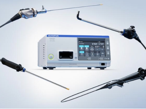
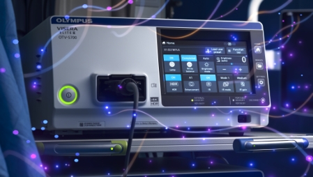
Created to accelerate procedures and learning curves for improved patient outcomes with advanced imaging, the VISERA ELITE™ III visualization system is a future-proof endoscopic visualization platform with software upgrades (e.g. NIR and 3D) and technology that allows you to focus on your procedures, while significantly reducing future costs.

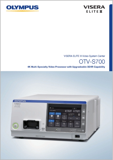
Designed to meet the needs of multiple specialties like general surgery, urology, gynecology, ENT and more, the VISERA ELITE III visualization system, offers 3D and 4K imaging, impressive fluorescence guided surgery and well proven, unique Narrow Band Imaging™ (NBI™), all in one system. Benefit from compatibility with existing scopes, camera heads and the latest generation* ENDOEYE™ series to efficiently reduce investment costs.
*Please contact Olympus for compatibility details.
Visualization Platform
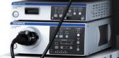
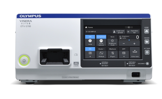
As a future-proof one for all system that grows with your individual needs, the VISERA ELITE III visualization platform is the endoscopic visualization platform with software upgrades designed to improve patient outcomes and standardize processes.
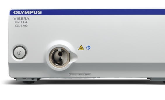
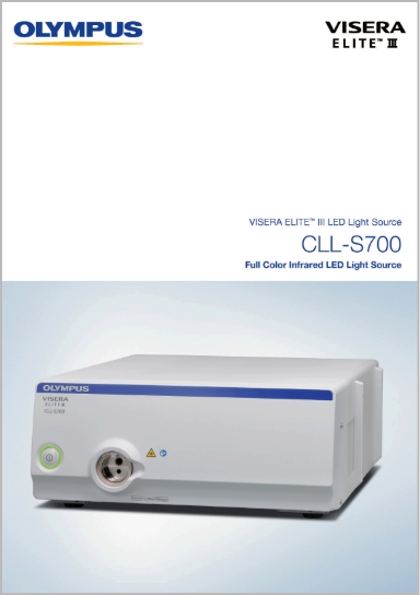
Experience unique natural full color reproduction with the VISERA ELITE III visualization system White Light IR LED Light source.
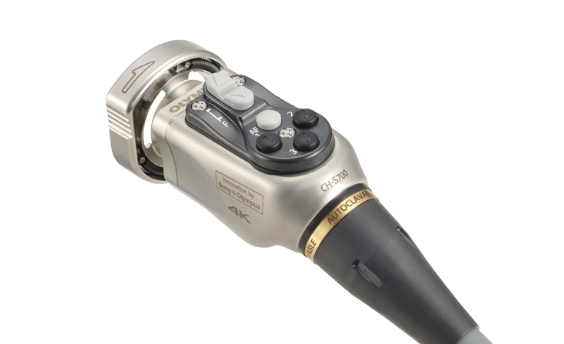
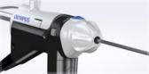
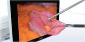
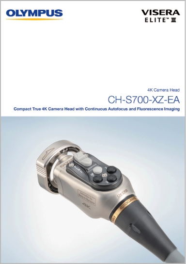
Discover incredibly fine details in true 4K image quality with the small and lightweight camera head featuring IR, NBI and YE observation modes. The new Continuous Auto Focus (CAF) and Extended Depth of Field (EDOF) features help to minimize distractions and broaden the focus area.
Benefit from 3D and 2D with a High Dynamic Range (HDR) setting to provide more natural contrast and reduced image saturation. The Advanced Image Multiple Enhancer (AIME) produces sharp, vivid image of structures without increasing noise.
The ENDOEYE HD II is a video laparoscope that combines vivid visualization and comfortable control. The advanced chip-on-the-tip technology provides bright and clear images.
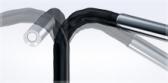
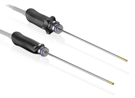
Easily shift the field of view with ENDOEYE Flex videoscope 5 mm and 10 mm by controlling the tip angulation up to 100° to the desired location. With even better access to narrow cavities, obtain the best viewing angle of the structures being visualized. In particular, observation performance is improved around the rectum, VATS, pelvic cavity, retroperitoneal approach to urology, etc.
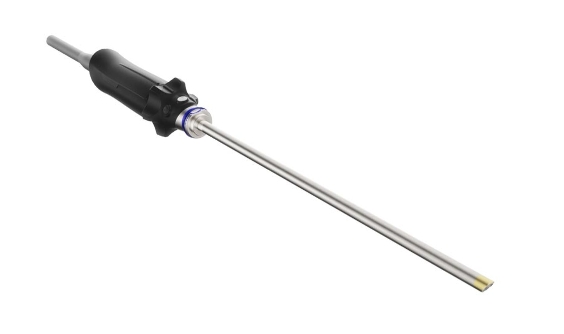
Perform 3D procedures without compromise. The autoclavable, rigid ENDOEYE 3D provides comfortable, natural 3D depth perception with a wide field of view and realistic colors.
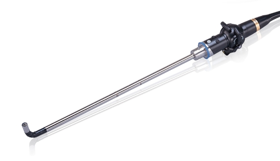
Change the direction of view while maintaining a stable horizon. ENDOEYE Rigid 30° supports your continuous critical view and provides you a reliable orientation even at challenging viewing angles with its continuous mechanical rotation function.

Enter the next generation of surgical imaging with scalable software upgrades now. No need to upgrade all your Olympus® equipment at once. You can maximize the utilization of existing, compatible components, benefit from a step-by-step approach and update as replacements are needed and future innovations are available over time.
Simply upgrade functionalities like 3D and IR and be ready for further visionary innovations via software activation.

VISERA ELITE III visualization system is your new versatile OR standard. Different disciplines can choose configurations and use peripheral technology according to individual needs. This provides standardization of OR workflows along with true flexibility. Only pay for what you need, keeping the possibility for future upgrades through software licensing.
Are you interested in experiencing the VISERA ELITE III visualization platform? Request more information or a live demonstration and an Olympus consultant will contact you.
Request InformationImproved Surgical Outcome
Identify anatomical structures like nerves, arteries, ureter and pleura that are surrounded by fatty tissue with the Yellow Enhancement (YE) mode.
Visualize the most minute vascular and mucosal patterns and with the help of Narrow Band Imaging Technology (NBI).
Focus on your procedures in a constant sharp picture through 4K CMOS and the unique Continuous Auto Focus (CAF) and Extended Depth of field (EDOF) functions.
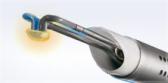
Discover impressive fluorescence guided surgery for better perfusion control and easier identification of biliary structures. Choose the IR gain that suits best and experience three selectable IR modes.
Medical Expert Training
Hands-on courses, e-learnings, workshops, peer-to-peer trainings, accredited continuing trainings, and custom on-demand learning for physicians who want to develop their skills and knowledge.
Browse through our lists and find the education that fits best to your needs.
Find Now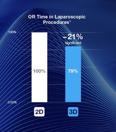
Benefit from realistic 3D vision and a high depth of field. The natural image allows laparoscopy to be performed much more precisely for even better patient outcomes.1 3D vision helps to save up to 35% and on average 21% operation time against standard 2D image procedures.2 Better usability can improve not only your surgical confidence but also your overall efficiency by helping to reduce OR time and accelerate patient recovery.2, 3, 4
Discover impressive fluorescence guided surgery for better perfusion control and easier identification of biliary structures, choose the IR gain that suits best and experience three selectable IR modes.
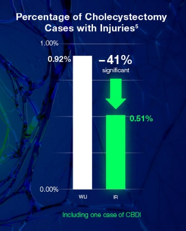
Minimize anastomotic leakages, better visualize vital structures and identify even more of the invisible in the future while potentially reducing post-operative complications by almost 50%.5 This helps to accelerate patient recovery and save total medical costs.
Complement the VISERA ELITE III visualization platform with a wider ecosystem of solutions. Complement the surgical visualization with innovative insufflation, full energy portfolio of advanced surgical devices and next level connectivity abilities – all from one source – for increased efficiency and high quality of surgery.
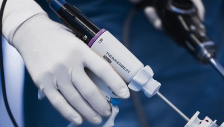
Benefit from combining pioneering surgical visualization and advanced surgical devices. Our surgical portfolio of instruments and generators provides the full range of specialized and unique devices and equips you with the power for your best performance.
The hospital-wide nCare™ medical recorder connected to our telecollaboration solution and our VaultStream™ medical content management solution makes it easy to view, record, stream, annotate, label and edit surgical video information pre-, intra- and post-operatively.
Are you interested in experiencing the VISERA ELITE III visualization platform? Request more information or a live demonstration and an Olympus consultant will contact you.
For more information, contact an Olympus Representative at 1-800-387-0437 / 1-289- 269-0100.
1. How Technology Can Impact Surgeon Performance: A Randomised Trial Comparing 3-Dimensional versus 2-Dimensional Laparoscopy in Gynecology Oncology. Fanfani F, Rossitto C, Restaino S, Ercoli A, Chiantera V, Monterossi G, Barbati G, Scambia G. J Minim Invasive Gynecol. 2016 Jul-Aug;23(5):810-7. doi: 10.1016/j.jmig.2016.03.020. Epub 2016 Apr 1. PMID: 2704674
2. Comparison of two- and three-dimensional display for performance of laparoscopic total gastrectomy for gastric cancer. Kanaji S, Suzuki S, Harada H, Nishi M, Yamamoto M, Matsuda T, Oshikiri T, Nakamura T, Fujino Y, Tominaga M, Kakeji Y. Langenbecks Arch Surg. 2017 May;402(3):493-500. doi: 10.1007/s00423-017-1574-9. Epub 2017 Mar 17. PMID: 28314905.
3. 3D VS. 2D-Imaging in Laparoscopic Procedures: Opportunity Costs Associated with the Reduction of Time in the Operating Room (OR). L. Bruno, A. Zervakis, P. Reinders. SURGERY - MEDICAL TECHNOLOGIES| VOLUME 23, SUPPLEMENT 2, S740, DECEMBER 01, 2020
4. Impact of Three-Dimensional Laparoscopy in a Bariatric Surgery Program: Influence in the Learning Curve. Padin EM, Santos RS, Fernández SG, Jimenez AB, Fernández SE, Dacosta EC, Duran AR, Artime Rial M, Dominguez Sanchez I. Obes Surg. 2017 Oct;27(10):2552-2556. doi: 10.1007/s11695-017-2687-5. PMID: 28456885.
5. Fluorescent cholangiography significantly improves patient outcomes for laparoscopic cholecystectomy. Broderick, R.C., Lee, A.M., Cheverie, J.N. et al. Surg Endosc (2020).